
Specialties
- Body Imaging
- MSK Imaging
- Neurology & Neuro-Imaging
- Cardiovascular Imaging
- Imaging Technology
- Informatics
CTIPM's advanced imaging techniques enable physicians to better diagnose and treat patients suffering from neurologic diseases such as Alzheimer's disease, primary and metastatic brain tumors, movement disorders, multiple sclerosis, and epilepsy.
3-Dimensional Image Acquisition and CTI Post Processing Allows Precise Tracking and Quantification of Neurodegeneration
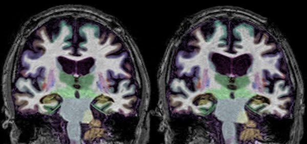
A 73 year old patient with memory complaints was imaged using CTI's Post-Processing for Quantitative Segmental Volume Assessment. At baseline, the patient showed lower-than-expected volumes of the brain's memory structures (Left image, in yellow). Two years later, the same patient was imaged again (Right image).
Providing quantification of interval change, CTI's precise image registration and segmentation also allows the radiologist to view the exact same slice position, regardless of head positioning, yielding improved visualization of change across time. As can be seen, the images provide objective support for an ongoing neurodegenerative process. The patient was subsequently enrolled in a clinical trial of a new Alzheimer's disease therapeutic.
Quantitative imaging, the practice of extracting quantifiable information from magnetic resonance imaging (MRI), as well as advanced MR imaging techniques can help physicians better diagnose and treat patients with various neurological diseases.
For example, in neurodegenerative conditions that impair memory, this approach to imaging can help physicians measure the location and severity of brain damage impacting memory, enabling them to more precisely diagnose and treat their patients. In the field of neuro-oncology, quantitative imaging as well as advanced diffusion weighted imaging (also known as Restriction Spectrum Imaging, or RSI-MRI), magnetic resonance spectroscopy (MRS), and magnetic resonance perfusion (MRP) help clinicians to provide more accurate diagnoses and prognoses and to better assess how patients are responding to treatments.
Neuro-Oncology:
Memory Loss:
Movement Disorders:
Multiple Sclerosis:
Epilepsy:
Developmental/Congenital Disorders:

James B. Brewer, MD, PhD
Chair of Neurosciences
Professor of Radiology at UC San Diego
Director for Neurology & Neuro-Imaging, CTIPM

Nikdokht (Niky) Farid, MD
Associate Professor of Radiology at UC San Diego
Director for Neurology & Neuro-Imaging, CTIPM
Neurodegenerative (or Memory Loss)
Movement Disorders
Multiple Sclerosis
Neuro-Oncology
Neurosurgery
Radiation Oncology